Ultrasound

Voxel Radiology has invested in state-of-the-art ultrasound machines( Philips EPIQ) . We believe our machines are world class providing advanced diagnostic imaging capabilities.
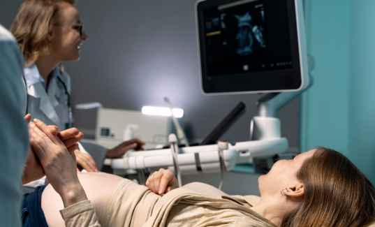
ultrasound

What is Ultrasound ?

An ultrasound is an imaging examination that uses high frequency sound waves to create pictures of various parts of the body. The reflected sound wave echoes are recorded and displayed as real-time visual images. Ultrasound uses no radiation and a technician called a Sonographer performs the scan.Ultrasound is especially used in obstetrics and gynaecology, assessing soft tissues such as muscles and tendons, and in investigation of the abdominal organs, amongst other things.

FAQ

Ultrasound is generally considered safe when used appropriately by trained professionals. However, like any medical procedure, it does carry some risks and considerations:
  1. Potential Biological Effects: Ultrasound energy can cause heating or mechanical effects in tissues. While diagnostic ultrasound uses low-intensity waves, prolonged exposure or improper use could potentially cause tissue damage. This risk is minimised through adherence to safety guidelines and limits on exposure time and intensity.

  2. Fetal Risks: Ultrasound is commonly used during pregnancy, but excessive or unnecessary exposure might pose risks to the developing fetus. Current guidelines recommend using ultrasound only when medically indicated during pregnancy to minimise potential harm.

  3. Misdiagnosis: Ultrasound images can sometimes be misinterpreted, leading to incorrect diagnoses or unnecessary medical interventions.

  4. Discomfort: In some cases, especially with certain types of ultrasound procedures, patients may experience discomfort or pain.

  5. Overuse: While ultrasound is valuable for diagnostic purposes, overuse or unnecessary examinations may expose patients to potential risks without corresponding benefits. It's important to note that these risks are generally low when ultrasound is performed by trained professionals following established guidelines. Pregnant women and others undergoing ultrasound examinations should discuss any concerns with their healthcare provider to ensure the procedure's appropriateness and safety.

Depending on the type of ultrasound scan you need, you may be asked to fast or drink a lot of water before your appointment. For example, for abdominal ultrasound, you might need to fast for several hours before the appointment. This will assist the sonographer to view certain areas. Our administration team will advise of any instructions to follow prior to an appointment.

For most ultrasound appointments, it's typically recommended to wear comfortable and loose-fitting clothing. Here are some specific guidelines depending on the type of ultrasound you're having:
  1. General Ultrasound (Abdominal, Pelvic, etc.):
    - Wear comfortable clothing that allows easy access to the area being examined. For example, for abdominal ultrasound, you might wear a loose-fitting shirt or blouse and pants or a skirt that can be easily pulled up.

  2. Transvaginal Ultrasound:
    - You may be asked to change into a gown provided by the clinic or hospital. It's helpful to wear easy-to-remove clothing, such as a skirt or loose pants.

  3. Breast Ultrasound:
    - Wear a top that can be easily removed or unbuttoned from the front, as you may need to expose the area for the ultrasound exam. A two-piece outfit can be convenient.

  4. Thyroid Ultrasound:
    - Wear a top that allows easy access to your neck area. You may be asked to remove any jewelry around your neck. In general, avoid wearing clothing with too many layers or tight-fitting garments that may restrict access to the area being examined. If you're unsure about what to wear, you can always call ahead and ask for guidance based on the type of ultrasound you'll be undergoing.

During an ultrasound appointment, the following typically happens: Preparation: Depending on the type of ultrasound, you may be asked to change into a gown to expose the area of interest that needs to be examined.

Procedure Explanation: The healthcare provider (usually a sonographer) will explain the procedure, including what to expect and any potential discomfort.

Gel Application: A water-based gel is applied to the skin over the area being examined. This helps transmit the ultrasound waves and ensures good contact between the skin and the ultrasound transducer.

Transducer Placement: The sonographer then places the ultrasound transducer, which is a handheld device that emits and receives ultrasound waves, on the skin and moves it around to obtain images of the internal organs or structures.

Image Capture: During the procedure, the sonographer captures images in real-time on a monitor. They may also take measurements or record specific images for the healthcare provider to review.

Post-Procedure: After the necessary images are obtained, the gel is wiped off, and you may be allowed to clean up and change back into your clothes.

Interpretation: The images are interpreted by a radiologist or healthcare provider who will then discuss the findings with you, either immediately or at a follow-up appointment. The entire procedure is typically painless, though you may experience slight discomfort from pressure as the transducer is moved over the skin. Ultrasound appointments are generally quick and non-invasive, with minimal preparation required depending on the type of ultrasound being performed.

There are different types of ultrasound scans available depending on the purpose and location to be examined. Here are some common types of ultrasound:
  1. Abdominal Ultrasound: This examines organs in the abdomen, such as the liver, gallbladder, pancreas, kidneys, and spleen.

  2. Pelvic Ultrasound: Used to examine the organs in the pelvic area, including the uterus, ovaries, and prostate.

  3. Obstetric Ultrasound: Specifically used during pregnancy to monitor the growth and development of the fetus.

  4. Transvaginal Ultrasound: A type of pelvic ultrasound where a small transducer is inserted into the vagina to obtain clearer images of the pelvic organs, especially useful for examining the uterus and ovaries.

  5. Breast Ultrasound: A type of pelvic ultrasound where a small transducer is inserted into the vagina to obtain clearer images of the pelvic organs, especially useful for examining the uterus and ovaries.

  6. Thyroid Ultrasound: Examines the thyroid gland for nodules, cysts, or enlargement.

  7. Musculoskeletal Ultrasound: Used to evaluate muscles, tendons, ligaments, joints, and soft tissues for injuries, inflammation, or abnormalities.

  8. Vascular Ultrasound: Examines blood flow and blood vessels using Doppler ultrasound. Each type of ultrasound serves a specific diagnostic purpose and may require different techniques or approaches to obtain clear images of the targeted area. The choice of ultrasound type depends on the symptoms, medical history, and the area of concern for the patient